|
作者:Desiree E. Morgan
作者单位:Department of Radiology, University ofAlabama at Birmingham, JTN452 619 South 19th Street, Birmingham, AL 35249, USA 本文发表于:AbdominalImaging & #40;2014& #41; 39:108–134 翻译:Shuai Zhang, M.D.

上期回顾: 能谱CT能够提高对胰腺富血供病变或囊性肿块复杂内部结构的清晰显示。碘基图有助于评价胰腺坏死,在急性胰腺炎的发作中对后腹膜的低密度物质集聚区进行分析,可以帮助判断是否存在胰腺自溶。对于肝脏或胰腺切除手术,可能会使用胆道金属支架或手术金属夹,这些金属物体会产生金属伪影,影响胰腺的观察。使用能谱CT的金属伪影去除序列(MARS)或采用140 keV图像都可以去除金属伪影,更好的观察胰腺。
本期内容: 单源瞬切能谱CT在胃肠道疾病诊断中的应用价值
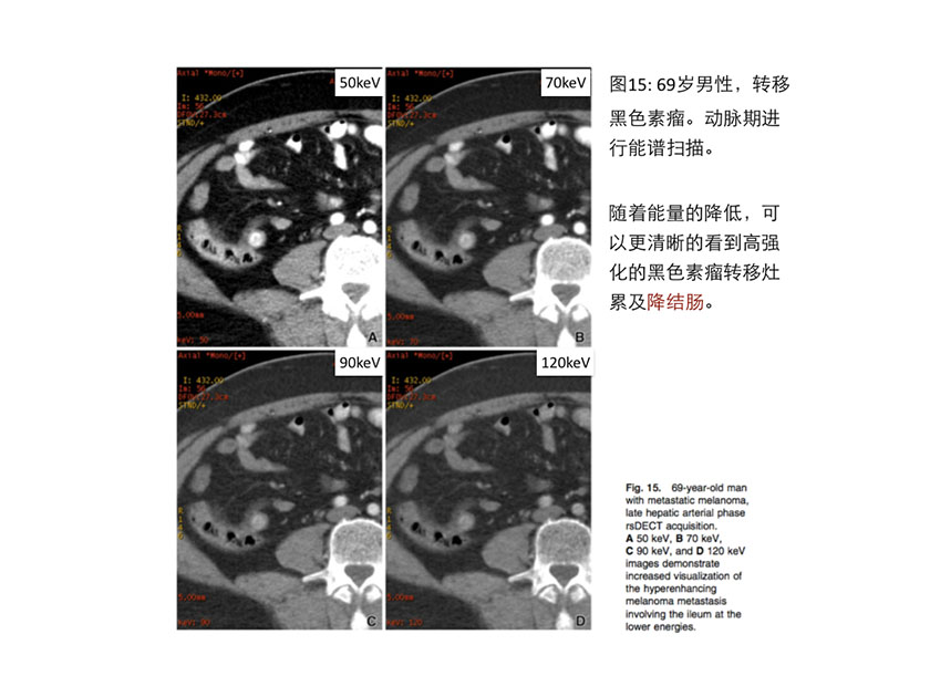
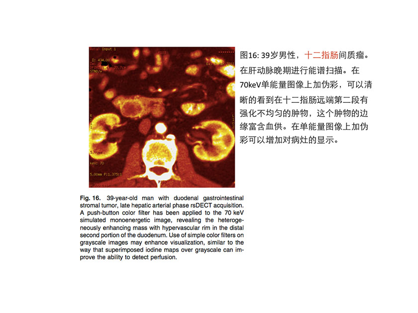
单源瞬切能谱CT在胃肠道的应用 Gastrointestinal tract applications 单源瞬切能谱CT在胃肠道的应用价值包括提高富血供肠道病变的显示效果,更好的诊断缺血性肠病,评估可替代对比剂的临床效果和改进CT结肠成像时“电子肠道清洁”的效果。 Potential benefits of utilizing dual-energy CT in the gut include possibilities forimproved visualisation of hyper-enhancing bowel pathologies, betteridentification of ischemic segments, evaluation of alternate contrast agents,or improved methods of electronic colon cleansing during CT colonography. 如果用CT来诊断活动性炎性肠病,低keV图像可以明显提高“高强化小肠段”的检出敏感性。(其原理和80kVp成像可提高Crohn’s病诊断效能类似 [55])。 Similar to evaluation of Crohn disease at 80 kVp [55]compared to 120 kVp, improved sensitivity to detect hyper-enhancing segments ofactive small bowel inflammation using lower keV viewing energies might bepossible, if CT rather than MRI is being utilized to establish the diagnosis. 同理,低keV单能量图像还可以提高黑色素瘤转移灶(见图15)和胃肠道神经内分泌肿瘤(见图16)的检出敏感性。 Identification of melanoma metastases & #40;Fig. 15& #41; or small gastroenteric neuroendocrineneoplasms & #40;Fig. 16& #41; may also benefit from low keV viewing or low kVpacquisition and viewing.


在已知或高度怀疑癌症的人群中,碘基图像可以帮助检出未做肠道准备的病人的结肠腺癌(中位直径4.3 cm)[56]。 Identification of colonic adenocarcinomas & #40;median size 4.3 cm& #41; in unprepped patients was aidedby the use of iodine maps in a population with known or highly suspectedcancers [56]. 两组科研人员对于胃肠道间质瘤转移的患者进行单源瞬切能谱成像,结果均显示相对于肿瘤大小缩小和Choi标准,能谱碘定量是一种更加稳定和敏感的疗效评估指标,用来评价靶向药物治疗的效果[57–59]。这种新方法也可用于其它富血供肿瘤的评估& #40;见图 17& #41;,碘浓度可以作为替代灌注的生物指标,其降低代表肿瘤对抗血管生成药物有反应。 The utility of dual-energy CT in patients with metastatic gastrointestinal stromal tumorshas been evaluated by two groups, both of whom suggested that iodinequantification rather than size de- crease and Choi criteria might be a morerobust indicator of response to therapy with targeted agents [57–59], and thistype of novel analysis can be translated to other hypervascular tumors & #40;Fig.17& #41;, where an iodine content decrease represents a surrogate biomarker forperfusion and thus response to anti-angiogenic agents.

最后,在用兔子进行的创伤模型的动物实验研究中,用单源瞬切能谱CT的基物质分离图像,有/无经验的阅片者都能对腹腔外渗液进行准确而自信的判断,鉴别外渗液是源于静脉对比剂外渗还是口服铋剂外渗。[60] Finally, in a rabbit model of trauma, experienced and inexperienced readers alike wereable to distinguish with increased confidence and accuracy extraluminalabdominal collections that arose from IV iodinated contrast extravasationcompared to those originating from oral Bismuth contrast extravasation, usingrsDECT material decomposition attenuation maps [60].
总结 Summary 能谱CT在腹部的应用正在不断的丰富: • 应用单能量图像可以更好的发现病灶和观察病灶。 • 同时单能量图像在低能量段可以提高碘的CT值(因为更加接近碘的K峰),因此利用单能量图像可以更少的应用碘对比剂。 • 应用基物质图像可以进行虚拟平扫、碘定量和结石成分定性分析。 • 基物质图像还可以扩展到对非碘类物质的定性和定量研究。 • 能谱CT可以有效的去除硬化伪影。 • 能谱CT还可以在腹部多期扫描中省去平扫序列,从而降低患者的辐射剂量。 The abdominal applications of dual-energy CT continue to evolve. • There is great potential for improved diagnosticperformance by capitalizing on increased lesion conspicuity with lower keVmonoenergetic images or lower kVp image sets. • Material-specific imaging allows generation of virtualunenhanced images, iodine quantification, and renal stone characterization atpresent, and • the possibilities exist to create new types of contrastor expand qualitative and quantitative tissue analyses for substances otherthan iodine. • Artifacts due to beam hardening and pseudo-enhancementin abdominal structures can be lessened with the rsDECT approach. • Because post-contrast rsDECT simulated monoenergetic HUvalues increase at the lower end of the energy spectra as the k edge of iodineis approached, just as with low kVp imaging the opportunities to use lesseramounts of iodinated contrast are being explored. • Finally, both clinically available technologies, dsDECTand rsDECT, can be utilized to reduce radiation dose by obviating the need forconventional unenhanced series from multi-phasic abdominal protocols.
《连载9》引用的参考文献: 55. Kaza R, Platt J, Al-Hawary M, et al. & #40;2012& #41; CT enterographyat 80 kVp with adaptive statistical iterative reconstruction versus at 120 kVpwith standard reconstruction: image quality, diagnostic ade- quacy, and dosereduction. AJR 198:1084–1092 56. Boellaard T, Henneman O, Streekstra G, et al. & #40;2013& #41; Thefeasi- bility of colorectal cancer detection using dual-energy computedtomography with iodine mapping. Clin Radiol 68& #40;8& #41;:799–806 57. Schramm N, Schlemmer M, Englhart E, et al. & #40;2011& #41; Dualenergy CT for monitoring targeted therapies in patients with advancedgastrointestinal stromal tumor: initial results. Curr Pharm Bio- technol 12:547–557 58. Apfaltrer P, Meyer M, Meier C, et al. & #40;2012& #41;Contrast-enhanced dual-energy CT of gastrointestinal stromal tumors. InvestigRadiol 47& #40;1& #41;:65–70 59. Meyer M, Hohenberger P, Apfaltrer P, et al. & #40;2013& #41; CT-basedre- sponse assessment of advanced gastrointestinal stromal tumor: dual energyCT provides a more predictive imaging biomarker of clinical benefit than RECISTor Choi criteria. Eur J Radiol 82& #40;6& #41;:923–928 60. Mongan J, Rathnayake S. Fu Y, et al. & #40;2013& #41; Extravasatedcontrast material in penetrating abdominopelvic trauma: dual-contrast dual-energy CT for improved diagnosis-preliminary results in an animal model.Radiology 268& #40;3& #41;:738–742
|