|
作者:Desiree E. Morgan 作者单位:Department of Radiology, University of Alabama at Birmingham, JTN452 619 South 19th Street, Birmingham, AL 35249, USA 本文发表于:Abdominal Imaging (2014) 39:108–134 
上期回顾:
CT双能量成像可以产生物质相关图像:双源双能量减影可以在图像空间进行物质减影,减掉图像中的碘,获得虚拟平扫图像。单源瞬切能谱成像可以在原始数据空间产生基物质图像,可以定量的分析NIST(美国国家技术与标准管理局)数据库的各种物质,例如碘、钙、铁、二氧化硅等。 本期内容: 单源瞬切能谱CT的单能量成像。

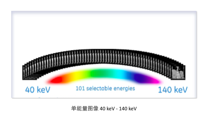
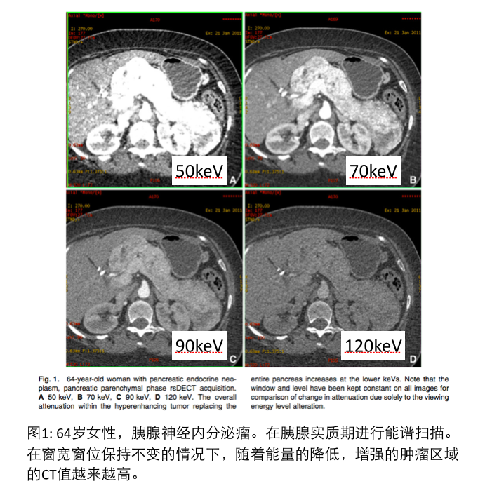
单源瞬切能谱CT可以利用80kVp和140kVp的原始数据,在“原始数据空间”(又名:投影数据空间)产生单能量图像(而不是产生传统的80kVp和140kVp的Dicom图像)。每一层面都可以产生从40keV 到 140keV 101个单能量图像,其中70keV或者78keV的图像被用来推送到PACS进行诊断。相对于传统的混合能量图像,单能量图像可以减少硬化伪影,进行更精准的CT值测量 [4]。最近的一个研究证实了单能量图像可以减少肾囊肿的假强化现象[5]。在专业能谱工作站上,医生可以和调节窗宽窗位一样,方便快捷的选择最适合诊断要求的单能量图像。 On the rsDECT system, simulated mono-energetic, also referred to as monochromatic images, are created in "projection" or "raw data space" from the detector data, without generation of actual 80 and 140 kVp images. From a practice standpoint, although 101 possible viewing energies exist for each image of the CT examination, images generated from either the 70 or 78 keV energy are sent to PACS and used for diagnosis. One advantage of simulated monochromatic images over low kVp images is that reduction in beam hardening artifacts is inherent, and more quantitatively accurate attenuation measurements may be obtained [4], a feature confirmed by reduction of renal cyst pseudo-enhancement in a recent phantom study [5]. Also with the rapid-switching system it is possible using either the dedicated workstation or thin client server to view simulated monoenergetic images that range from 40 to 140 keV dynamically or on-the-fly, much as one would to window and level a standard CT image, selecting the energy that best depicts disease processes.

因为碘的K峰是 33.2 keV,因此随着X线能量的降低,图像中碘的增强效果就会越来越明显(参见图1)。 Because the k edge of iodine is 33.2 keV, just below the lower end of the scale of spectral imaging available with the rsDECT system, the contribution of iodine to attenuation of the images increases as the viewing keV level decreases (Fig. 1).

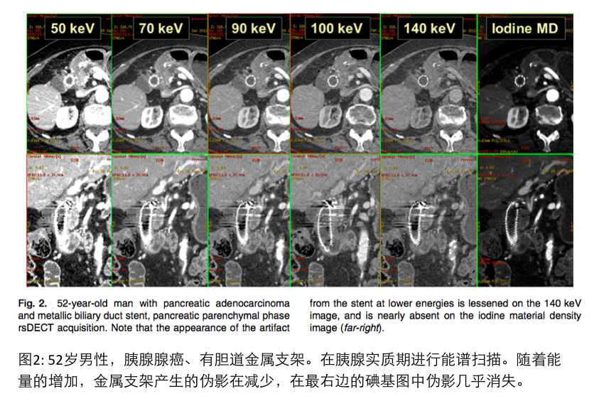
因此,在临床增强扫描中,50-52keV的单能量图像更适合观察胰腺[7]和肝脏的病灶,110-140keV的单能量图像可以减少金属伪影 [8](参见图2)。在2012年美国腹部影像学年会上,Weber在大会演讲中描述了一个现象:140keV的单能量图像可以最大程度的消除碘的增强效应,产生肾上腺的“虚拟平扫”图像,有助于诊断肾上腺富含脂肪成分的腺瘤。 It has become apparent in our practice that viewing iodinated IV contrast enhanced rsDECT abdominal images at 50–52 keV consistently better depicts lesion contrast in the pancreas [7] and liver, whereas viewing abdominal images at 110– 140 keV lessens metal [8] artifacts (Fig. 2); viewing at 140 keV maximally removes (but not completely) the effects of iodine on the monoenergetic images thereby producing ‘‘pseudo-unenhanced’’ images of the adrenal gland which may help to confirm the presence of a lipid-rich adenoma (Weber et al. (2012) presented to the annual meeting of the Society of Abdominal Radiology, unpublished data).

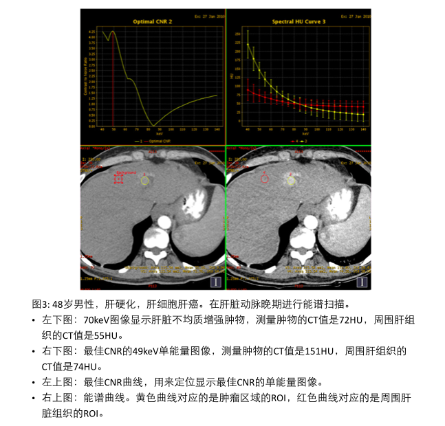
随着能量的降低,单能量图像中的噪音也随之增加。为了平衡组织对比度和噪音之间的关系,专业的能谱后处理工作站具备“最佳对比噪声比CNR”功能。通过这个功能,医生可以方便的找到对比噪声比(CNR)最佳的单能量图像,用来提高病灶的探查效率。(参见图3)
Simulated monoenergetic image noise also increases as the keV decreases, similar to the noise increase on 80 kVp images compared to 120 kVp. The optimal balance between tissue contrast and noise can be calculated with a push button feature on the independent workstation of the rsDECT scanner, and a ‘‘CNR-optimized’’ level can then be viewed to enhance lesion conspicuity (Fig. 3).
《连载3》引用的参考文献: 4. Yu L, Leng S, McCollough CH (2012) Dual-energy CT-based monochromatic imaging. AJR 199:S9–S15 5. Jung DC, Oh YT, Kim MD, et al. (2012) Usefulness of the virtual monochromatic image in dual-energy spectral CT for decreasing renal cyst pseudoenhancement: a phantom study. AJR 199(6): 1316–1319 6. Matsumoto K, Jinzaki M, Tanami Y, et al. (2011) Virtual mono- chromatic spectral imaging with fast kilovoltage switching: Im- proved image quality as compared with that obtained with conventional 120-kVp CT. Radiology 259(1):257–262 7. Patel BN, Thomas JV, Lockhart ME, Berland LL, Morgan DE (2013) Single-source dual-energy spectral multidetector CT of pancreatic adenocarcinoma: optimization of energy level viewing significantly increases lesion contrast. Clin Radiol 68(2):148–154 8. Pessis E, Campagna R, Sverzut JM, et al. (2013) Metal artifacts reduction using monochromatic images from spectral CT: evalua- tion of pedicle screws in patients with scoliosis. Eur J Radiol . doi: 10.1016/j.ejrad.2013.02.024
下期预告: CT双能量成像中的CT值和辐射剂量。
|