|
作者:Desiree E. Morgan 作者单位:Department of Radiology, University of Alabama at Birmingham, JTN452 619 South 19th Street, Birmingham, AL 35249, USA 本文发表于:Abdominal Imaging (2014) 39:108–134 
上期回顾: 本综述从硬件技术和临床应用的角度阐述了CT双能量成像在腹部各器官应用的最新进展。从硬件平台上讲,CT双能量成像分为双源双能量减影和单源高低压瞬切能谱CT。双源双能量减影从图像空间减影,产生融合和减影图像;能谱CT从原始数据空间进行分析,产生基物质图像和单能量图像。 本期内容: CT双能量物质相关图像:减影和基物质图像
相对于传统CT,双能CT可以产生本质上完全不同的图像。 Dual-energy CT scanners are capable of making fundamentally different kinds of images. 一、双源双能量减影可以在图像空间产生物质减影图像。 dsDECT can generate in images space the material subtraction images. 双源双能量减影可以实现“物质减影”。“物质减影”基于一个假设:腹部的每个像素点都由软组织、碘和脂肪组成。根据这个假设,“物质减影”可以计算出每一个像素点碘的分布,然后通过减影方法把碘减掉,产生虚拟平扫的图像。 With dsDECT, the material subtraction is a three-point material subtraction process where it is assumed that every voxel in the abdomen is composed of soft tissue, iodine, and fat, and a post-processing algorithm generates a map that encodes the iodine distribution in each voxel, then subtracts it from the image.

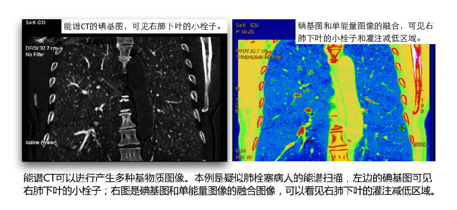
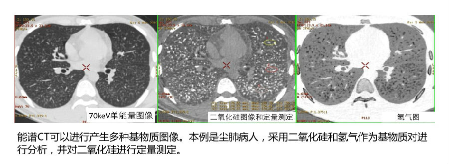
二、能谱CT(单源高低压瞬时切换技术)可以产生基物质图像。 rsDECT can generate material density images for diagnosis. 能谱CT基物质图像基于基本的物理原理:特定元素(例如钙、碘、金、钆)或者特定物质(例如脂肪、血液、粘液等)会针对X线的不同能量产生不同的线性衰减系数。利用这个特性,我们可以通过能谱CT来区分这些物质。 One type of image is the material density image, sometimes referred to as material-specific image. Because linear attenuation coefficients obtained at different X-ray energies are unique for a given element, such as calcium, iodine, gold, gadolinium, or a substance such as fat, blood, mucin, etc, the change in attenuation between the two energy spectra used for DECT can be used to discrimi- nate the materials. 
通过计算每个像素点中特定物质的含量,能谱CT可以产生诸如碘基图、尿酸图、虚拟平扫图等基物质图像。 This computational process which determines the amount of a substance within each image voxel is used to produce clinically relevant presentations of the data such as iodine images or overlays, uric acid maps, or a virtual unenhanced image, sometimes referred to as virtual noncontrast image. 
能谱CT的基物质图像是通过“基物质解析“过程实现。这种过程可以分析水/碘、钙/碘、脂肪/碘等。因为这种方法的灵活性,能谱CT可以定量分析 NIST(美国国家技术与标准管理局)物质数据库中具备的各种物质(例如:钆、金、铁、血液、粘液等)。 With rsDECT, this process uses material decomposition basis pairs of water(–iodine) and iodine(–water) to create the virtual unenhanced and iodine material images. On the rsDECT workstation, the same material decomposition basis pair process can evaluate other substances, such as calcium(–iodine) and iodine(–calcium), or fat(–iodine) and iodine(–fat); any material for which a NIST (National Institute of Standards and Technology) attenuation curve is available (gadolinium, gold, iron, blood, mucin, etc) can be installed into the system and then the material can be rapidly evaluated quantitatively on the images. 
《连载2》引用的参考文献: 1. Megibow AJ, Sahani D (2012) Best practice: implementation and use of abdominal dual-energy CT in routine patient care. AJR 199:71–77 2. Primak A, Ramirez G, Liu X, et al. (2009) Improved dual energy material discrimination for dual-source CT by means of additional spectral filtration. Med Phys 36(4):1359–1369 3. Graser A, Johnson T, Chandarana H, et al. (2009) Dual energy CT: preliminary observations and potential clinical applications in the abdomen. Eur Radiol 19:13–23 下期预告: 单源瞬切能谱CT的单能量图像。
|