|
1-Beat:狭窄精确诊断
Detailed and Reliable Stenosis Detection in One Heart Beat
作者:Ronny R. Buechel, M.D.(苏黎世大学核医学科,瑞士)
翻译:郭勇 刘华阳 审校:任心爽 吕滨(中国医学科学院阜外心血管医院)
病史 Patient History 60岁男性,短暂性脑缺血发作& #40;TIA& #41;后典型心绞痛。运动负荷试验临床症状阳性,心电图结果阴性。行冠状动脉CT检查。 A man in his 60s complaining of typical angina pectoris and status after TIA was referred to coronary CT angiography after an exercise test clinically positive and electrically negative.
Revolution CT 采集参数 1-beat 心脏成像(图1) 160mm轴扫模式,心电门控 100kv,270mA 75%期相采集 体重指数25 ASiR-V迭代重建技术 旋转速度:0.28s/rot 心率:46-48 BPM DLP: 45 mGy-cm 有效辐射剂量:0.63mSv

图1: 1-beat 心脏采集
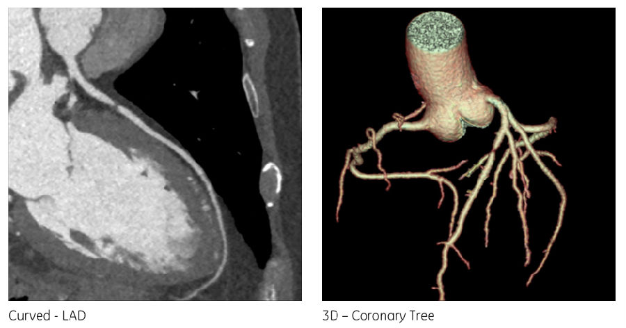
结论 该患者钙化积分为0,但是在CT影像上可以看到左前降支一处90-99%的重度狭窄(图2)。 In this patient with a calcium score of 0, a subtotalocclusion of the LAD & #40;90-99 %& #41; could be visualized on the CT images.
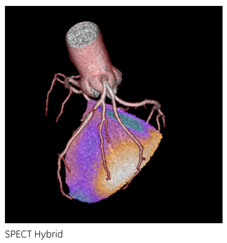
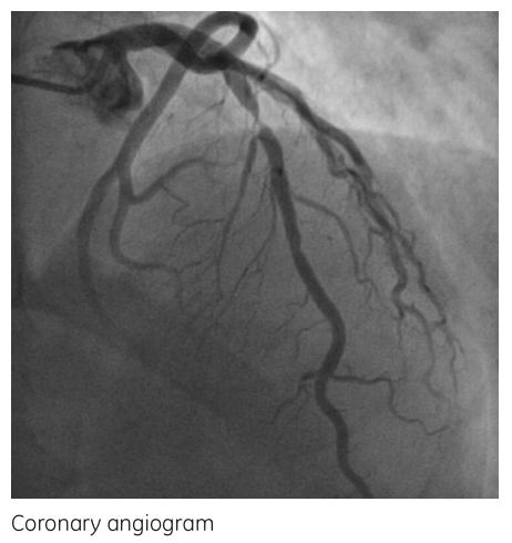
SPECT显示在相应的前壁有一个可逆性灌注缺损区(图3)。该患者进行了冠状动脉造影并确定诊断(图4),之后进行了支架置入术。 Correspondingly, SPECT revealed a large reversible perfusion defect in the anterior wall. The patient underwent coronary angiogramwhere the diagnosis was confirmed and stenting of the lesion was performed. 
图2: CCTA 图像 
图3: SPECT和CCTA的融合图像 
图4: 冠脉造影图像
Ronny R.Buechel 医生的评价 “由于一个心动周期的图像采集和新的迭代重建技术ASiR-V 的应用,Revolution CT 可以在低kV的条件下得到高质量的心脏影像。Revolution CT在超低剂量的条件下,展示了优异的软斑块鉴别能力。” Thanks to the one-beat axial acquisition, the new iterative reconstruction technology ASiR-V and the highimage quality at low kV, Revolution CT delivers cardiac CT with excellent softplaque differentiation at very low dose in routine use.
|