|
作者:Desiree E. Morgan 作者单位:Department of Radiology, University of Alabama at Birmingham, JTN452 619 South 19th Street, Birmingham, AL 35249, USA 本文发表于:Abdominal Imaging & #40;2014& #41; 39:108–134
翻译:Liu, Shichen, M.D. 
上期回顾: 有了CT能谱成像之后,即使不进行传统的CT平扫,也能通过能谱的虚拟平扫图像来确定肾上腺病变的性质。能谱特征性的水基图、脂肪基图和140keV单能量图像可以提供确诊富脂质肾上腺腺瘤的的诊断阈值,这个阈值具备很高的特异性。
本期内容: 能谱CT在肝脏疾病诊断中的应用价值
能谱CT在肝脏病诊断中的应用价值 Liver applications
能谱CT在肝脏的应用包括通过基物质图像的形态学观察和定量测量以及低keV单能量图像的形态学观察,增加肝脏病灶的检出。 Evolving liver applications include material-specific image viewing and quantification as well as low keV monoenergetic image viewing to increase lesion conspicuity. 基物质图像可用于评估胆道结石、肝实质内脂肪或铁沉积;同时定量分析肿瘤内碘的存在,这样不仅可以检出肿瘤病灶,同时可以评估治疗后的疗效。在100名胆囊或者胆管结石的研究中,双源双能减影( dsDECT )的肝动脉晚期或者门静脉期的虚拟平扫图像可以导致16%的胆结石漏诊,而这些结石在传统的平扫中可以被发现。漏诊的主要是小于1.7mm的胆囊结石以及CT值在78HU左右的色素性胆管结石[42]。 Material-specific images may be used to evaluate biliary stone disease, hepatic parenchymal deposition of fat or iron, or identification of iodine within tumors both as a means of detection of disease as well as assessment of response. Virtual unenhanced images generated on a dsDECT system from late hepatic arterial and portal venous phase failed to identify 16% of gallstones seen on conventional unenhanced images in a study of 100 patients with gallbladder or bile duct stones. These were predominantly small gallstones & #40;<1.7 mm& #41; and pigmented bile duct stones with HU<=78 HU [42]. 在临床实践中,当肝脏中出现铁元素的时候,使用传统CT去测量肝实质中的脂肪含量是非常困难的,反之亦然。也就是说,当肝实质内有铁存在时,脂肪含量会被低估;肝脏含有脂肪时,铁的含量也会被低估。CT双能量成像可以定量测定肝脂肪含量。Fischer在一个双能CT体模研究中,采用”三物质分离”算法,生成虚拟的“铁物质图像”,在不考虑肝实质和脂肪的情况下,可以准确测量铁的含量[43],同时生成虚拟的“不含铁的物质图像”,在有铁的存在下,准确测量脂肪含量[44]。 In clinical practice, it is difficult on SECT to quantify hepatic fat in the presence of iron and vice versa. The amount of fat is underestimated in the presence of iron, and the amount of iron is underestimated due to the presence of hepatic fat. In phantom studies using DECT and an iron-specific three material decomposition algorithm, Fischer et al. [43] were able to create a virtual iron image to accurately quantify iron irrespective of liver tissue attenuation and fat, and to generate a virtual non-iron image to accurately quantify fat in the presence of iron [44].
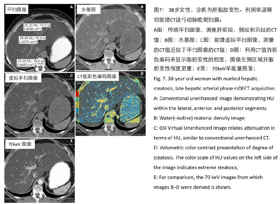
Joe等人进行了一个结合了体模和临床的研究,利用140kVp图像和80kVp图像间的平均密度差异,可区分含铁大于10%的肝脏,但是该结果和肝脂肪变性没有相关性 [45] 。请注意,上述研究的CT双能量数据都来自于平扫图像。 In a combined phantom and clinical study, Joe et al. [45] showed that the difference of averaged attenuation between the 140 and 80 kVp images could be used to differentiate patients with greater than 10% hepatic iron, but the same value did not correlate with hepatic steatosis. Note that the dual-energy acquisition for this study was without IV contrast enhancement. 在一个体模及小鼠肝脂肪变性的研究中,Artz等人对肝脏进行能谱平扫,体模研究显示肝实质密度及肝脂肪含量与甘油三酯含量高度相关;但在小鼠模型中,只有肝实质密度与肝细胞内甘油三酯的含量高度相关[46]。Zheng等学者应用一种创新的后处理方法,能够鉴别出人群脂肪沉积的不同程度[47]。 In a phantom and mouse model of hepatic steatosis, Artz et al. [46] demonstrated that attenuation and fat density of the liver parenchyma using unenhanced rsDECT correlated highly with triglyceride content in the phantom, but in the model, only attenuation correlated highly with triglyceride content in hepatocytes. Using novel post-processing of rsDECT monoenergetic image subtraction & #40;termed DESI& #41; in a patient population, Zheng et al. [47] were able to identify varying degrees of fat accumulation. 请注意,上述这些研究都是用双能CT进行平扫,并没有尝试对强化后的肝脏进行肝脂肪的定量分析。考虑到脂肪肝的高发病率,利用增强扫描的能谱数据就可以精确测量肝的脂肪含量(见图7),这对患者非常重要,因为患者就不用再增加一次平扫或者再去进行核磁共振的肝脏脂肪定量的检查。 Note that these studies did not attempt to address quantification of hepatic steatosis in a post-contrast setting. Given the prevalence of fatty liver disease, it will be important to use material-specific imaging capabilities to quantify hepatic fat after administration of IV contrast for patients who did not undergo an unenhanced SECT series & #40;Fig. 7& #41;. This is important, as the quantitative information of hepatic fat content can be gained from the images already acquired as part of the clinically indicated scan rather than having to suggest that the patient undergo a second unenhanced CT or an MRI for further evaluation. 
另外能谱CT的基物质图像,可以帮助探查到肝细胞肝癌的肿瘤栓子中的碘元素。应用癌栓的碘与主动脉碘的比值,Qian等研究者利用单源瞬切能谱CT可靠地区分出肿瘤栓子与血栓 [48]。 Another application explored with material-specific imaging of the liver is the identification of iodine within tumor thrombus in patients with hepatocellular carcinoma; using iodine indices & #40;comparing thrombus to aorta& #41; Qian et al. [48] reliably distinguished bland from tumor thrombus in patients using rsDECT. 利用单源瞬切能谱CT的单能量图像(低keV)或者双源双能减影CT80kVp的图像都可以提高肝脏病灶的对比噪声比。尽管Altenbernd等研究者发现使用双源CT的动脉期80kVp图像可以提高肝脏富血供病灶发现的敏感性,但是80kVp图像主观评分的图像质量非常差。另外该研究使用的早期双源CT,40个病例中,15个肝脏无法全部覆盖[49]。建议应用一种新的图像融合算法可以提高图像质量[50]。 Lesion contrast and conspicuity in hepatocellular carcinomas may be improved by viewing lower keV images on rsDECT or the low kVp images on dsDECT. However, although Altenbernd et al. found better sensitivity for detection of hyperenhancing liver lesions with arterial-phase 80 kVp images compared to blended images on dsDECT, the subjective image quality of the 80 kVp images was judged very poor. In addition, 15 of 40 subjects in that study had incomplete hepatic coverage with the early generation dsDECT machine [49]. Applying novel blending parameters might help to improve image quality [50]. 使用单源瞬切能谱CT扫描,可以利用一键式最佳CNR技术,迅速在40-140keV的图像范围内定位出最利于病灶观察的单能量keV图像,用于肝脏及腹部其他脏器的病变的观察(见图3)。Thomas 等研究者发现,使用这一技术来比较最佳keV图像与70keV图像,大量肝脏局限性病灶在动脉期被发现而且不会降低图像质量。 进一步的大样本、多评估者比较的大型研究正在进行中。 Using the rsDECT system, it is possible to instantaneously calculate the optimal viewing keV & #40;from a range of 40–140& #41; for a lesion within the liver or other abdominal organs, based on contrast to noise ratios & #40;CNR& #41; of the image & #40;See Fig. 3& #41;. Using this technique and comparing the CNR-optimized keV image to the 70 keV image, a greater number of focal hepatic lesions were identified on arterial-phase rsDECT without compromising image quality & #40;Thomas et al. & #40;2011& #41; presented to the American Roentgen Ray Society, unpublished data& #41;; further study with a larger multi-reader protocol is ongoing.


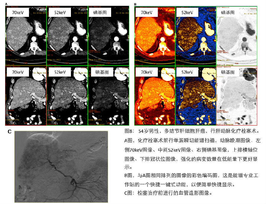
如图8所示,使用单源瞬切能谱的专业分析平台,可以在一个显示器窗口中并列对比观察低keV图像、70keV图像及碘基图像。实践表明,碘基图像同样可以增加病灶检出。彩色融合图像(见图8)、碘基图像、能谱HU曲线,结合方便单能量图像浏览方法,可以动态观察所有能量,以展示各种能量信息。 When images are viewed with dedicated rsDECT software, it is possible to view the lower keV images and 70 keV images side by side with iodine images & #40;Fig. 8& #41;, a practice which also enhances lesion detection due to the lower CNR of the iodine images. Color coding & #40;see Fig. 8& #41;, iodine threshold imaging, spectral HU curves, and a slide-bar that allows on the fly scrolling through all available energies provide additional means of presentation the dual-energy information.
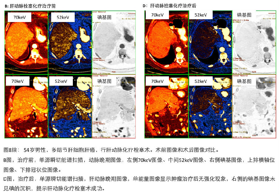
单源瞬切能谱CT还有助于便捷的评估肝脏病变局部治疗的治疗效果,例如肝动脉化疗栓塞术或者射频消融治疗;也可应用碘的定量测量来评估疗效(参见图9) [51, 52]。 同双能量减影图像相比,能谱的碘基图像因为具备更高的对比噪声比,可提供更好的消融治疗的病灶边缘显示 [52]。 This can be helpful in rapidly visualizing response to locoregional therapies such as transarterterial chemoembolization or ablation; alternatively, quantitative measures can be utilized & #40;Fig. 9& #41; [51, 52]. Higher contrast to noise ratios of iodine maps provide improved conspicuity of ablation zone margins compared to blended images [52]. 
《连载6》引用的参考文献: 42. Kim JE, Lee JM, Baek JH, et al. & #40;2012& #41; Initial assessment of dual- energy CT in patients with gallstones or bile duct stones: can virtual nonenhanced images replace true nonenhanced images? AJR 198:817–824 43. Fischer MA, Reiner CS, Raptis D, et al. & #40;2011& #41; Quantification of liver iron content with CT—added value of dual-energy. Eur Radiol 21:1727–1732 44. Fischer MA, Gnannt R, Raptis D, et al. & #40;2011& #41; Quantification of liver fat in the presence of iron and iodine. Investig Radiol 46& #40;6& #41;:351–358 45. Joe E, Kim S, Lee KB, et al. & #40;2012& #41; Feasibility and accuracy of dual-source dual-energy CT for noninvasive determination of he- patic iron accumulation. Radiology 262& #40;1& #41;:126–135 46. Artz NS, Hines C, Brunner ST, et al. & #40;2012& #41; Quantification of he- patic steatosis with dual-energy computed tomography. Investig Radiol 47& #40;10& #41;:603–610 47. Zheng X, Ren Y, Phillips WT, et al. & #40;2013& #41; Assessment of hepatic fatty infiltration using spectral computed tomography imaging: a pilot study. J Comput Assist Tomogr 37& #40;2& #41;:134–141 48. Qian LJ, Zhu J, Zhuang ZZG, et al. & #40;2012& #41; Differentiation of neoplastic from bland macroscopic portal vein thrombi using dual- energy spectral CT imaging: a pilot study. Eur Radiol 22:2178–2185 49. Altenbernd J, Heusner TA, Ringelstein A, et al. & #40;2011& #41; Dual-en- ergy-CT of hypervascular liver lesions in patients with HCC: investigation of image quality and sensitivity. Eur Radiol 21:738–743 50. Wang Q, Shi G, Liu X, et al. & #40;2013& #41; Optimal contrast of computed tomography portal venography using dual-energy computed tomography. J Comput Assist Tomogr 37& #40;2& #41;:142–148 51. Lee JA, Jeong WK, Kim Y, et al. & #40;2013& #41; Dual-energy CT to detect recurrent HCC after TACE: initial experience of color-coded iodine CT imaging. Eur J Radiol 82& #40;4& #41;:569–576 52. Lee SH, Lee JM, Kim KW, et al. & #40;2011& #41; Dual-energy computed tomography to assess tumor response to hepatic radiofrequency ablation. Investig Radiol 46& #40;2& #41;:77–84
|
共 1 个关于综述:CT双能量成像在腹部的应用(连载七) Part 7:能谱CT在肝脏疾病诊断中的应用价值,每天更新的回复 最后回复于 2014-08-12 15:05:41