|
综述:CT血管造影(CTA)20年的发展之路(连载六):CT血管造影术对于临床的贡献:冠脉疾病的CT血管造影
作者:Geoffrey D. Rubin, MD 作者单位:Duke Clinical Research Institute, 2400 Pratt St 本文发表于:Radiology:Volume 271: Number 3—June 2014 radiology.rsna.org 翻译:张杰;校对:张帅、陆伟;整理:梁军

上期回顾: 本综述从以下两个方面:1)主动脉腔内支架植入术 2)经导管主动脉瓣植入术(TAVI),来探讨CT血管造影术对于主动脉腔内修复术方面的临床应用和价值。
本期内容: 短短20年的时间,CT血管造影已从一个新兴的成像方式,演变为一个关键的临床工具,在体内几乎所有血管性疾病的诊断和治疗中起着主导的作用。本期内容着重从冠脉疾病的CT血管造影,从临床价值、对特异性心肌缺血、辐射剂量等方面进行总结和探讨。
冠脉疾病的CT血管造影:最后的前沿 CT Angiographyof Coronary Artery Disease: The Final Frontier
自1990年代以来,CT技术的快速发展实现了几乎所有血管的CT血管造影应用程序。然而,一个最重要的动脉---冠状动脉的CT血管造影依然无法开展。由于位于心脏的表面,冠状动脉一直不断地在运动。提高CT血管造影时间分辨率的策略就是要“冻结”心脏运动,并且能够清楚地显示冠脉及邻近的心脏结构。 Through the 1990s, the rapid evolution of CT technology provided a means for CT angiography applications to develop for virtually every vascular bed. However,one of the most important arterial beds, the coronary arteries, remainedelusive. Owing to their position on the surface of the heart, the coronary arteriesare in constant motion. Strategies to improve the temporal resolution of CT angiography were needed to "freeze" cardiac motionand allow clear depiction of the coronary arteries and adjacent cardiacstructures.
虽然CT冠脉造影可以追溯到1990年代中期使用前瞻性心电门控触发扫描的电子束CT& #40;84& #41;,但除了一些研究中心之外其有限的应用阻碍了电子束CT技术的发展。当多排探测器CT在1998年首次推出时,其时间分辨率远比100 –msec的电子束CT低得多。尽管降低CT床速使得回顾性心电门控的全心动周期采集叠加投影成像大大拓宽了冠脉CT血管造影的临床应用(27),然而对于四排多层螺旋CT而言,回顾性门控的实用性还是有限的。 While coronary CT angiography dates back to the mid-1990s using the ECG to prospectively trigger electron-beam CT & #40;84& #41;, the limited availability of electron-beam CT prevented the technique from developing beyond a few centers. When multi-detector CT was first introduced in 1998 its temporal resolution was substantially worse than the 100-msec resolution of electron-beam CT. Strategies that slowed the CT table to allow retrospective, ECG-gated binning of projectionsacquired throughout the cardiac cycle substantially broadened the availability of coronary CT angiography & #40;27& #41;, however with four-row multi-detector CT,retrospective gating was of limited practicality.
4年中,16排多层螺旋CT的出现和更快的机架旋转大幅度地提高了冠脉CT血管造影 & #40;85& #41;,它随着CT技术的每一次进步而继续发展和提高。今天,冠脉CT血管造影的不断发展,确立其在怀疑冠脉疾病(CAD)诊断检查中的作用(图11)。 Within 4 years, 16-row multi-detector CT and faster gantry rotations brought substantially greater robustness to coronary CT angiography & #40;85& #41;, which has continued to evolve and improve with each iteration of CT technology. Today,coronary CT angiography continues to evolve and establish its role in the diagnostic workup of suspected coronary artery disease & #40;CAD& #41; & #40;Fig 11& #41;.

图11:63岁,男性,高血压和高脂血症,呈现弥漫性胸部疼痛和气急。2年前的常规血管造影发现尚正常。 (a)心电门控CT血管造影显示的曲面重建显示左前降支(LAD)中段广泛非钙化斑块,造成严重的狭窄(箭头)。 (b)由覆盖整个心动周期的1 0组序列重建获得的局部心肌室壁运动的彩色编码图可映射冠脉病变与前壁和左室心尖部运动机能的减退的心功能的结果(紫色)(箭头)。 (C)冠脉病变(箭头)在随后的常规血管造影被证实。
在患者已知或怀疑有CAD的背景下,通过CT血管造影评价解剖已一再证明CAD无创性评估的整体高效性和无可匹敌的阴性预测值(86-88)。在一个包括了28项研究,涉及1286例患者的多元分析中,采用与现在稍有点过时的,64排螺旋CT技术检查冠脉狭窄50%或以上的冠脉CT血管造影,并使用常规冠脉造影为参考标准的研究,该研究报告了患者的敏感度为99%,特异性为89%,阳性预测值93%,阴性预测值为100%& #40;89& #41;。每支血管分析的阴性预测依然是100%。 Within the context of patients known to have or suspected of having CAD, anatomic evaluation by means of CT angiography has repeatedly demonstrated overall high performance and an unmatched negative predictive value for non-invasive assessment of CAD & #40;86-88& #41; . Ameta-analysis of 28 studies, involving 1286 patients, investigating the performance of coronary CT angiography with the now somewhat dated technology of 64-row multi-detector CT for the detection of coronary stenosis of 50% orgreater, using conventional coronary angiography for a reference standard, reported a per-patient sensitivity of 99%, specificity of 89%, positivepredictive value of 93%, and a negative predictive value of 100% & #40;89& #41;. Negative predictive value remained 100% on per-vessel analyses.
在当前对于疑似稳定性心绞痛的指导原则中,CT血管造影术已被指定为在大多数情况下适宜于轻到中度的、之前为非诊断的或模棱两可的检查结果的患者(92)。比照慢性胸痛,急性胸痛的诊断方法更不确定。只有一小部分患者有足够明确的体征和症状证明直接推荐采用常规血管造影& #40;93& #41;,而大多数患者被归为中等风险,定义为30%---70%的有意义的CAD预测概率& #40;92& #41;。对于这些患者,需要额外的检查来避免在急诊或胸痛诊所长时间的留观所致的风险和费用。CT血管造影俨然成为一种最有效的方法可靠地排除这些疾病& #40;93 -95& #41;。 Within current guidelines for the evaluation of suspected stable angina, CT angiography has been designated appropriate for low-to-intermediate-like lihood patients with prior non-diagnostic or equivocal test results as the most common scenario & #40;92& #41;. In contrast to chronic chest pain, the diagnostic algorithm for acute chest pain is much more volatile. Only a small percentage of patients present with sufficiently definitive signs and symptoms to justify direct referral to conventional angiography & #40;93& #41;, while a majority of patients are classified as being at intermediate risk, defined as 30%-70% pretestprobability of significant CAD & #40;92& #41;. For these patients, additional testing is needed toavoid the expense of prolonged stays in emergency departments or chest pain units. CT angiography appears to be emerging as one ofthe most efficient and effective methods for reliably ruling out disease& #40;93-95& #41;.
两个主要的多中心随机临床试验,涉及1370例和1000例急性胸痛患者的CT血管造影与传统的护理的对比,发现在急诊科和短期急诊留观者有明显较高的直接出院率,而无主要心血管不良事件发生率的增加(93,94)。 Two major multicenter randomized trials involving 1370 and 1000 patients with acute chest pain compared the use of CT angiography to traditional care and found significantly higher direct discharge rates from the emergency department and shorter emergency department stays without an increase in the rate of major adverse cardiac events & #40;93, 94& #41;.
而证据支持冠脉CT血管造影的临床常规使用目前高于任何其他的CT血管造影应用,且疑似CAD患者此类应用的数量也高于采用其他所有CT血管造影者,对CT血管造影广泛用于胸痛患者的评估所致的成本问题的过度担忧已导致在美国高度多样化的补偿和冠脉CT血管造影的利用。 While the level of evidence in support of the routine clinical use of coronary CT angiography is higher currently than for any other application of CT angiography and the number of patients who present with suspected CAD dwarfs that of allother CT angiography indications, heightened concerns over the cost of widespread CT angiography utilization in the evaluation of patients with chestpain has resulted in highly heterogeneous reimbursement and thus utilization of coronary CT angiography in the United States.
这是5年后的两组关于主要不良心脏事件的低中度风险患者分析分别表明有26%-33%的CAD相关费用的减少而无不良心血管事件或与CAD相关的住院治疗的增加,接着初期评价了CT血管造影与单光子发射CT(SPECT),一线心功能评价的传统方式(95,96)。 This remains 5 years after two analyses of patients at low and intermediate risk of major adverse cardiac events, respectively, demonstrated a 26%-33% reduction in CAD-related costs without an increase in adverse cardiovascular events or CAD-related hospitalization, following initial evaluation at CT angiography when compared with initial evaluation at single photon emission computed tomography & #40;SPECT& #41;, the traditional modality for first-line cardiac function assessment & #40;95, 96& #41;.
对于冠脉CT血管造影表现的一个重要关注在于其阳性预测值不如其阴性预测值那样完美准确。鉴于鉴别诊断中CAD临床症状的高发病率,依然担心, 常规使用冠脉CT血管造影所致的假阳性结果将增加下游成本。 One important concern regarding the performance of coronary CT angiography is that its positive predictive value is not nearly as robust as its negative predictive value.Given the high prevalence of clinical symptoms where CAD is within the differential diagnosis, there remain concerns that false-positive results from the routine use of coronary CT angiography will increase downstream costs.
两种新型方法正在出现即可以通过功能信息与标准的形态学评价CT心肌灌注显像和CT血流储备分数(FFRCT)将大幅度提高冠脉CT造影的阳性预测值。前者的技术已经通过CT技术的不断进步而实现,而后者的技术要依赖于复杂的后处理及冠脉CT血管造影数据的分析。 Two strategies are emerging that could substantially improve the positive predicative value of coronary CT angiography by associating functional information with the standard morphologic assessment-CT imaging of myocardial perfusion and CT-derived fractional flow reserve & #40;FFR ct& #41; measurements. The former technique has been enabled through ongoing advances in CT technology, while the latter technique relies on sophisticated post-processing and analysis of coronary CT angiographic data.
心肌灌注 Myocardial Perfusion
在当前的临床实践中,冠脉病变的血流动力学相关性正是源自于利用SPECT,正电子发射断层扫描术& #40;PET& #41;的心肌灌注,及至最近,心脏MR灌注成像,为冠心病的风险分层,指导治疗策略和结果预测提供了方法& #40;97& #41;。 In current clinical practice, the hemodynamic relevance of coronary lesions hasbeen derived from measures of myocardial perfusion by using SPECT, positrone mission tomography & #40;PET& #41; , and more recently, cardiac perfusion MR imaging, offering a means for risk stratification, guidance for treatment strategy, and outcome prediction & #40;97& #41;.
最初CT洞察到的心肌血供来源于诊断,“静态”的冠脉CT血管造影检查。心肌衰减模式被用来定性地评估心肌的血容量,作为心肌灌注的一项指标在首过动脉期。低密度区域伴随着相关冠脉的狭窄预示着有意义的CAD血流动力学的改变(98)。然而,这种方法的重大的局限性是近似的心肌灌注测量包括在扫描时可变的正常心肌强化及无法去量化结果。 Initial CT-derived insights into the myocardial blood supply have been derived from diagnostic, "static" coronary CT angiographic examinations. Myocardial attenuation patterns are used to assess myocardial blood volume qualitatively, as a surrogate for myocardial perfusion during the first-pass arterial phase. Hypoattenuating territories accompanied by stenosis within associated coronaries are indicative of hemodynamically significant CAD& #40;98& #41;. However, important limitations of this approach to approximate myocardial perfusion measurements include the variability of normal myocardial enhancement during image acquisition and an inability to quantify the results.
最近CT技术的两个进步使得基于CT的心肌灌注测量变得更加完美。先前文章中谈到在下肢动脉血流评估中消除管壁钙化影响的话题,双能CT有这个潜力来解决心肌内的碘浓度问题& #40;99& #41;。这种方法能够改进心肌血供的CT评价结果,其与MR及SPECT灌注成像& #40;100& #41;相对照而大幅增加相应的解剖信息。双能CT也已被证明能够改进图像质量和减少伪影如射线硬化等在CT灌注成像方面的传统局限性& #40;101& #41;。在双能技术用于改善心肌灌注评价中,当在心肌灌注的首过动脉期中被用于单期项采集的一部分时,它的定量能力是有限的& #40;98& #41;。
Two recent advances in CT technology are enabling more robust CT-based measurement of myocardial perfusion. Discussed previously within the context of minimizing the impact of mural calcium on the assessment of the lower extremity arterial runoff, dual-energy CT has the potential to resolve iodine concentration within the myocardium & #40;99& #41;. This method can improve the CT assessment of myocardial blood supply yielding results that are comparable to MR imaging and SPECT perfusion imaging & #40;100& #41; but augmented substantially by the associated anatomic information. Dual-energy CT has also been shown to allow for improvements in image quality and reductionin artifacts such as beam hardening that have traditionally limited CT perfusion imaging& #40;101& #41;. While dual-energy technology appears to improve myocardial assessment, when used as a part of auni-phasic acquisition during the first arterial pass through the myocardium, its capabilities for quantification are limited & #40;98& #41; .
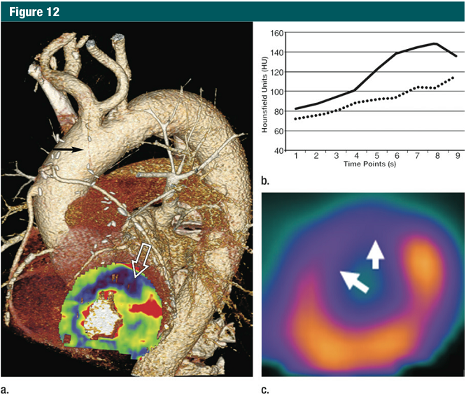
这后面的问题可以通过第二个进步来解决,即当对比剂流经心肌时提供宽探测器和快速穿梭容积成像,从而得到动态CT心肌灌注成像& #40;图12& #41;。动态碘对比的CT灌注能提供一个绝对定量测定心肌血流量,它优于MR(心肌灌注),后者的心肌信号与钆浓度之间呈非线性关系& #40;102& #41;。 This latter concern may be eliminated through the second advance that provides wide area detectors and the ability to rapidly shuttle across the imaging volume during the transit of contrast material through the myocardium, thus enabling dynamic CT myocardial perfusion imaging & #40;Fig 12& #41;. Dynamic iodine-based CT perfusion can provide an absolute quantitative measure of myocardial blood flow, an advantage over MR imaging, which suffers from a non-linearity in the relationship between myocardial signal and gadolinium concentration & #40;102& #41;.

图12:71岁,男性,冠心病史,三支冠脉搭桥术后,复发心绞痛。 (a)药物负荷试验,具备时间分辨率的CT心肌灌注图与冠脉CT血管造影解剖图叠加显示移植到左前降支(黑色箭头)的左内乳动脉闭塞,与之关联的负荷诱导的灌注缺失的在左室前壁心肌(白开箭头)。 (b)健康的(实线)和病变的心肌(虚线)在整个CT灌注持续时间内的衰减值(时间密度曲线)显示缺血心肌有持续的轻度强化。 (c)药物负荷SPECT研究呈现的灌注缺失与CT灌注结果有很好的关联,在相同的位置(箭头)。
动态CT灌注测量研究支持阈值在75-90ml˙100ml-1˙min-1,低于该值冠脉病变应考虑有血液动力学意义& #40;103- 105& #41;。对动态CT灌注的一个担忧是与大范围采集相关的辐射剂量的增加;但是,据报道辐射剂量是类似于SPECT灌注成像& #40;105& #41;,即获取灌注信息的传统方法。 Investigations of dynamic CT measurements of perfusion support a threshold of 75-90 mL-100 mL-1.min-1,below which coronary lesions should be considered hemodynamically significant& #40;103-105& #41;. One concern related to the use of dynamic CT measurements of perfusion is the increased radiation dose associated with the extended acquisition; however, the radiation doses are reported to be similar to that of SPECT perfusion imaging & #40;105& #41;, the traditional method for obtaining such information.
此外,随后将要讨论的,迭代重建方法的进步对于CT将进一步减少辐射。 Moreover, as will be discussed subsequently, advancements in iterative reconstruction methods for CT should result in further reductions in radiation exposure.
然而用CT评价心肌灌注的这些成果目前还相对处于初期阶段,但是将单一设备用于心脏缺血疾病检查,获得全面的解剖和功能评价,其吸引力还是无比巨大的。正当撰写这篇文章的时候,来自众多的单和多中心测试进行中的临床性能数据正期待着CT灌注在CAD诊断方法中所起的作用。 While efforts at deriving measures of myocardial perfusion with CT are currently in astate of relative infancy, the appeal of obtaining comprehensive anatomic and functional assessments in the workup of ischemic heart disease with a single instrumental set-up is strong. Atthe time of this writing, clinical performance data are pending from amultitude of ongoing single and multi center trials to establish the role of CT perfusion in the diagnostic algorithm of CAD.
从静息CT血管造影中估计病变特异性缺血 Estimating Lesion-Specific ischemia from Resting CT Angiography
以往稳定型心绞痛的治疗专注于通过造影测量血管直径减少的解剖狭窄血管的重建。最近的数据表明由侵入性测量血流储备分数(FFR)确定的病变特异性缺血所导向的血管重建可提高无病生存率,高于仅根据血管造影术& #40;106& #41;。 Historical management of stable angina has been focused on the revascularization of anatomic stenosis with an angiographic measure of diameter reduction being the guide. Recentdata has suggested that revascularization guided by lesion-specific ischemia as determined by invasively measured fractional flow reserve & #40;FFR& #41; improvesevent-free survival over decisions based on angiography alone & #40;106& #41;.
冠状动脉FFR是以一比值来衡量,即以药物诱导血流最大化后,冠状动脉狭窄远端的收缩压除以升主动脉收缩压。已经证明目测乃至定量评估血管狭窄与传统的造影或冠脉CT血管造影一起对于病变特异性缺血可提供有限的信息& #40;106,107& #41;。计算流体力学的进展和基于图像的建模以及冠脉CT血管造影图像质量的持续改进,已可以从常规的静息冠脉CT血管造影的图像来计算冠脉血流和压力(108,109)即允许FFR的计算而无需修改成像协议,增加图像,增加射线照射,或添加药物应激剂。 Coronary arterial FFR is measured as the ratio of the systolic blood pressure distal toa coronary artery stenosis divided by the systolic blood pressure in the ascending aorta during pharmacologically induced flow maximization. It has also been well established that visual inspection and even quantitative assessment of diameter reduction with either conventional or coronary CT angiography provide limited information regarding lesion specific ischemia & #40;106,107& #41;. Advances in computational fluid dynamics and image-based modeling as well as continued improvement in image quality of coronary CT angiography has allowed the calculation ofcoronary flow and pressure from a typically acquired routine resting coronary CT angiogram & #40;108,109& #41; allowing the calculation of an FFR without the modification of imaging protocols, additional imaging, increase in radiation exposure, or the addition of pharmacologic stress agents.
冠脉的血流量和压力可采用流体力学调节方程式(Navier-Stokes)推导获得,但直至最近依然没有计算能力或合适的非侵入性的冠脉模型被使用来推算无创的FFR。更完整的方法关于详述FFRct推导的基础超出了本文的范围,并已在其他文章有详细的描述& #40;110& #41;。 Coronary artery flow and pressure can be derived by using the governing & #40;Navier-Stokes& #41; equations of fluid dynamics, but until recently there has not been the computational power or an adequate noninvasive model of the coronary arteries to apply them to derive a non-invasive FFR. More complete methods describing in detail the basis for FFRct derivation are beyond the scope ofthis review and have been described in detail elsewhere & #40;110& #41;.
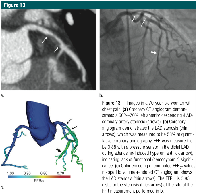
而只有一个新的进展,有越来越多的证据表明,以侵入性FFR为参考标准,高性能的FFRCT对于病变特异性缺血诊断有高的诊断准确性(图13)。最重要的,所有直至现在发布的数据显示FFRCT比单一的冠脉CT血管造影具有优势。 While only a recent development, there is increasing evidence for the high diagnostic performance of FFRct for the determination of lesion-specific ischemia, with invasive FFR as the reference standard & #40;Fig 13& #41;. Of importance, all published data until now have shown the superiority of FFRCT ascompared with coronary CT angiography alone.

图13:70岁,女性,伴胸痛。 (a)冠脉CT血管造影显示50% -70%左前降支(LAD)狭窄(箭头)。 (b)冠脉造影显示LAD狭窄(细箭头),它测得的狭窄在定量冠脉造影为58%。FFR测值为0.88,伴随LAD远端的负荷传导为腺苷诱导充血(粗箭头),表明缺乏功能(血流动力学)意义。 (C)FFRCT计算值的彩色编码映射到CT血管造影VR(容积再现)图上显示LAD狭窄(细箭头)。FFRCT值为0.85于狭窄远端(粗箭头)在FFR实际测量位置见b图。
一项前瞻性,多中心的国际试验发现,FFRct在所有诊断测量方面的表现已超越CT血管造影而成为缺血的预报者(108)。对于病变特异性缺血,FFRCT比CT血管造影显示有更高的识别能力(曲线下面积:0.90FFR CT vs 0.75冠脉CT血管造影,P=0.001)。研究人员还发现,CT血管造影显示的传统的解剖狭窄特征在FFRCT上只极少被加得上。 A prospective, multicenter international trial found that FFRct surpassed CT angiography as a predictor of ischemia across all measures of diagnostic performance& #40;108& #41;. FFR CT displayed significantly higher discriminatory ability than CT angiography for lesion-specific ischemia & #40;area under the curve: 0.90 for FFRc-r vs 0.75 for coronary CT angiography, P=.001& #41;. The investigators also found that traditional anatomic stenosis characterization at CT angiography added little to FFRct.
此外,一个明显的临床挑战,58%的患者通过定量冠脉血管造影至少有一支为40%-69%狭窄,其中47%的病变与经FFR发现的缺血相关。对于这些中等度冠脉狭窄,FFRCT展示了诊断准确性,敏感性,特异性,阳性预测值,和阴性预测值分别为86%,90%,83%,82%,和91%,比对的标准CT血管造影相同各项测值分别为56%,90%,26%,52%,和75% & #40;111& #41;。 Moreover, for the distinctly clinically challenging 58% of patients with at least one 40%-69% stenosis by means of quantitative coronary angiography, 47% of the lesions we reassociated with ischemia by means of FFR. For these intermediate stenosis, FFRCT demonstrated a diagnostic accuracy,sensitivity, specificity, positive predictive value, and negative predictive value of 86%, 90%, 83%, 82% , and 91%, respectively, compared with 56%, 90%, 26%, 52%, and 75% for the same measures derived by means of standard CT angiographic interpretation & #40;111& #41;.
最近,一个更大规模的试验证实,在逐一病例分析中,对于冠脉狭窄50%或更高所致缺血预测FFRct优于传统CT血管造影,显示更好的病变识别率& #40;曲线下面积:0.81和0.68,P =0.0002& #41;& #40;109& #41;。当一个决策规则被用一项分析:只有2毫米或更大的血管被包含在其中且所有血管狭窄小于30%被认为是非缺血性时,FFRct的表现是非常令人满意的,准确率达到了92%,而以CT血管造影为基础的鉴别血管狭窄的准确率为68%(P<0.0001)。 More recently, a larger trial confirmed that on a per-patient analysis, FFRct is superior to traditional CT angiography analysis for predicting ischemia instenosis of 50% or greater, showing significantly better lesion discrimination when compared with CT stenosis of 50% or greater & #40;AUC: 0.81 versus 0.68. P =.0002& #41; & #40;109& #41;. When a decision rule was applied where in only vessels 2mm or greater in size were included in the analysis and all vessels with lessthan 30% stenosis were considered non-ischemic, the performance of FFRct was extremely robust, with an accuracy of 92% for FFRct versus 68% for CT angiography-based stenosis characterization alone & #40;P < .0001& #41;
而当最后主要的应用进入到临床邻域,结合了CT制造商非凡的技术发展,图像处理和分析的进步,以及来自于放射学和心脏病学领域研究人员的奉献精神和创造力使得用于冠心病(CAD)的CT血管造影术经历了其应用层面的最为深刻的转变。基于迄今为止获得的知识和进一步创新的轨迹,冠脉CT血管造影有望不久的将来在CAD的诊断和治疗中将发挥重大作用。 While last among major applications to enter into the clinical realm, a combination of remarkable technologic developments by CT manufacturers, advances in image processing and analysis, and dedication and creativity of investigators from the fields of radiology and cardiology has enabled CT angiography of CAD to undergo the most profound transformation ofany CT angiography application. Based on the knowledge gained to date and the trajectory of further innovation, coronary CT angiography appears poised to play a major role in the diagnosis and management of CAD in the near future.
|