|
综述:CT血管造影(CTA) 20年的发展之路(连载四):CT血管造影术对于临床的贡献:外周血管疾病——CTA在扫描覆盖范围和重建图像可视化的突破
作者:Geoffrey D. Rubin, MD 作者单位:Duke Clinical Research Institute, 2400Pratt St 本文发表于:Radiology: Volume 271: Number 3—June 2014 radiology.rsna.org 翻译:郑颖;校对:张帅、陆伟;整理:梁军

上期回顾: 本综述介绍了CT血管造影术对于临床的贡献:急性主动脉综合症(AAS)——新的知识重新定义疾病分类。CTA不仅彻底改变了对AAS的诊断与治疗,而当扫描采集与心电图(ECG)同步时,则从根本上更易理解AAS的症状,它正成为对AAS进行诊断、定性、制定治疗计划的一项优越技术。 本期内容: CT血管造影术对于临床的贡献:外周血管疾病: CT血管造影在扫描覆盖范围和重建图像可视化的突破 随着多排探测器CT的应用,整个下肢动脉,包括从腹股沟上方流入动脉至腹股沟下方流经动脉的CT血管造影成为了可能
外周血管疾病: CT血管造影在扫描覆盖范围和重建图像可视化的突破 Peripheral Vascular Disease: Pushing the Envelope for CT Angiography Coverage and Visualization
随着多排探测器CT的应用,整个下肢动脉,包括从腹股沟上方流入动脉至腹股沟下方流经动脉的CT血管造影成为了可能(25)。最初受限于需要1分钟的扫描,2-3mm的层厚,而现在64排CT使用亚毫米级别的层厚,也只需要10秒钟就能完成扫描,尽管无需如此之快的扫描速度。 CT angiography of the entire lower extremity arterial system—which includes suprainguinal inflow vessels and infrainguinal runoff—became possible with the introduction of multi-detector CT& #40;25& #41;. While initially limited to1-minute acquisitions and 2–3-mm-thick sections, modern 64-row CT scanners canachieve 10-second acquisitions with submillimeter section thickness, al-thoughsuch fast scan times are not necessary.
下肢动脉血管造影(CTA)的纵向扫描范围通常是从胸12椎体水平到足底,扫描野25-750px,以保证足够的层面内分辨率,层面间分辨率(Z轴分辨率)通常是0.7-1.25mm(38)。虽然采用当前最先进的CT扫描设备,动脉血管造影(CTA)图像数据的采集是相对直接而简单,而与血管内对比剂到达保持同步,需要特别注意病变的下肢动脉,对比剂团注经过可能会延迟(39)。 The anatomic coverage for a lower extremity CT angiogram typically extends from T12 through the toes, with a 25–30-cm field of view, to provide adequate in-plane resolution & #40;38& #41;.Through-plane resolution & #40;in the z-axis& #41; is typically 0.7–1.25 mm. While lower extremity CT angiography data acquisition is relatively straightforward withstate-of-the- art equipment, synchronization with contrast medium delivery requires particular attention to the potentially delayed bolus propagation in adiseased lower extremity arterial tree & #40;39& #41;.
一个简单的解决方案是特意将扫描时间延长到40秒,并且可以预设一个可选的从膝关节上缘水平到足尖水平的附加扫描程序(39)。 A simple strategy is to deliberately acquire the CT data in a relatively long scan time of 40 seconds and preprograman optional second pass from above the knees down through the toes & #40;39& #41;.
随着多排螺旋CT的发展,外周血管CT血管造影需要先进的图像处理工作站,以适应由1000-2000幅CT横断面重建图像组成的数据组(40)。因为观察横断面原始图像既不切实际又无法充分与治疗医师全面地信息交流,所以下肢动脉CTA的一个重要挑战就是可视化和后处理。 Together with the advances of multi-detectorCT, peripheral vascular CT angiography necessitated advances in the capabilities of image processing workstations to accommodate datasets composedof 1000-2000 primary trans-verse CT reconstructions& #40;40& #41;. Because viewing transverse source images is both impractical and inadequate for communicating findings to treating physicians, an important challenge of lower extremity CT angiography is visualization and post-processing.
传统的3D血管成像技术,如最大密度投影(MIP)和容积再现(Volume Rendering),对于外周动脉疾病的患者常常会受限,因为近一半病人有严重的血管壁钙化或内置支架。这些妨碍了相应血管在诊断上与治疗上的可视化(41)。 The classic 3D techniques for displaying cardiovascular CT data, such as maximum intensity projection & #40;MIP& #41;and volume-rendered images are often limited in patients with peripheral arterydisease, because approximately half of these patients have significant vessel wall calcifications or stents in place. Both preclude visualization of the diagnostically and therapeutically relevant flow channels & #40;41& #41;.
血管管腔面的分析最好由曲面重建来完成,可以是每个动脉分支分别重建(25,42)或是多支血管的曲面重建(43)。下肢动脉图像的后处理工作比较耗时,要在大量容积检查数据中获得标准化的下肢动脉树图像,可由指定的专门技术人员或一个集中的3D实验室来完成。 Analysis of the vessel lumen is best achieved with curved planar reformation, either through each arterial branch separately & #40;25,42& #41; or as multipath curved planar reformations& #40;43& #41;. Processing of lower extremity CT angiography may be time-consuming,and practices with large examination volumes might benefit from using dedicatedtechnologists or a centralized 3D laboratory to generate standardized images ofthe lower extremity arterial tree.
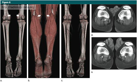
下肢CT血管造影(与磁共振血管造影)因为其广泛的适应症(38),最重要的是指导外周动脉疾病患者的治疗计划& #40;44& #41;,事实上已经取代了诊断性动脉内造影。其他适应症还包括急性下肢动脉缺血,创伤及解剖成像,如之前的游离皮瓣移植术(45),或疑有功能性或解剖性的运动员腘动脉受压综合征(图6)及髂动脉内纤维化(38)。 Lower-extremity CT angiography & #40;and MR angiography& #41; has virtually replaced diagnostic intra-arterial angiography for abroad spectrum of indications & #40;38& #41;, the most important of which is treatment planning for patients with peripheral artery disease & #40;44& #41;. Other indications include acute ischemia, trauma, andanatomic imaging, such as before free-flap harvesting & #40;45& #41;, or inathletes suspected of having functional or anatomic popliteal entrapmentsyndrome & #40;Fig 6& #41;, or iliac endofibrosis & #40;38& #41;.

图6 :19岁,篮球运动员,在球场上跑步的时候腿部出现疼痛 CT血管造影诊断为腘动脉受压综合征 (a)跖屈应力体位(负荷状态),VR图像显示,双侧腘动脉闭塞(箭头所示),同时小腿远段动脉几乎没有显示。(b)VR使用相同的数据、运用多层次交互显示方法显示下肢肌肉,腘动脉闭塞的位置(箭头所示)相当于腓肠肌内侧头部的水平,和图a相同(c)VR-CT血管造影示静息状态下、中立位,腘动脉和小腿动脉的正常图像。(d)负荷状态下,腓肠肌内侧头部水平的双侧异常腘动脉(箭头所示)(e)静息状态下,腓肠肌内侧头部水平的双侧腘动脉
在外周动脉病变的情况下,下肢CT血管造影的目的并不是进行确诊——诊断通常基于患者的症状、临床检查以及无创性检查如踝-肱指数,进行综合评价,其优势和意义在于呈现病变的程度和累及范围,指导治疗方案的选择。
The clinical role of lower extremity CT angiography in the setting of peripheral artery disease is not to establishthe diagnosis—this is typically based on symptoms, clinical examination, and noninvasive testing such as ankle-brachial index. The strength and role of lower extremity CT angiography is to map the disease process within this large territory, which is critical for treatment planning.
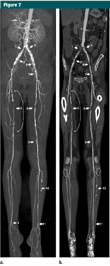
外周动脉疾病中需要实施临床干预的主要是两类疾病:患者生活受限的重度间歇性跛行(Fontaine IIb级)和重度下肢缺血(静息痛和/或组织缺损,Fontaine III级和IV级)(图7)(46)。对于跛行患者,治疗的目的主要是缓解症状,且通常局限于膝关节以上的病变。对于重度下肢缺血的患者,要进行血运重建,从而预防组织缺损和截肢,这需要对膝关节上下的血管进行积极的血管内治疗和/或外科手术的干预。 The two main categories of peripheral artery disease warranting intervention are patientswith lifestyle-limiting intermittent claudication & #40;Fontaine IIb& #41; and patients with critical limb ischemia & #40;rest-pain and/or tissue loss, Fontaine III and IV& #41;& #40;Fig 7& #41;& #40;46& #41;. In the claudication group, treatment aims at symptomrelief and is typically restricted to lesions above the knee. In patients withcritical limb ischemia, revascularization aims at prevention of tissue loss andamputation, which requires more aggressive endovascular and/or surgical treatment of arteries both above and below the knees.

图7:右下肢动脉缺血,并脚趾坏疽。 (a)最大密度投影(MIP) (b)多路径曲面重组(MPCPR)视图,下肢CT血管造影示双侧下肢弥漫性动脉粥样硬化性病变。髂总动脉75px范围的闭塞,导致右下肢的血供严重损害(1),髂外动脉长度为50px的重度狭窄(2),股浅动脉近中段狭窄(曲线箭头所指的长约125px的闭塞)(3),右侧胫后动脉起始部位发生闭塞,腓动脉在踝关节以上部位进行了代偿性血运重建(4),导致跨越踝关节处出现两支供血动脉原因。髂总动脉起始段75%的狭窄,导致左下肢血供受损(5),其造成的血运障碍重于髂外动脉的90%以上的狭窄(6,7)股浅动脉中段75px长的闭塞(8),股浅动脉远端中等程度的狭窄(9),左侧胫前动脉自起始部位至远端的250px范围的闭塞(10),腓动脉在踝关节以上水平进行了代偿性血运重建(11),使得跨越踝关节处出现两支供血动脉。 在MIP图像上被血管壁钙化所掩盖的病变(1,2,5,6,7),而在MPCPR视图上没有被钙化所掩盖,显示清晰。由于MPCPR图像是沿着血管中心进行重建,此时所呈现的非血管结构可能并不是标准的解剖关系。比如,在MPCPR图像中,腘动脉一侧的卵圆形高密度结构,代表的是股骨外侧髁和胫骨平台的侧面结构。
最近的一项综合分析报导:对于间歇性跛行患者,下肢动脉CT血管造影非常精确,敏感性达95%(95%可信区间:92%,97%),特异性96%(95%可信区间 93%,97%),而且能够探测到50%以上的狭窄和闭塞(47)。MR血管造影被报告有相似的敏感性(92%-99.5%)和特异性(64%-99%)(48)。 Lower extremity CT angiography is anaccurate imaging modality in patients with intermittent claudication, with asensitivity of 95% & #40;95% confidence interval: 92%, 97%& #41; and a specificity of 96%& #40;95% confidence interval: 93%, 97%& #41; for detecting more than 50% stenosis orocclusions reported in a recent meta-analysis & #40;47& #41;. Similarsensitivities & #40;92%–99.5%& #41; and specificities & #40;64%– 99%& #41; have been reported for MR angiography & #40;48& #41;.
在MR血管造影和CT血管造影这两种技术间的一项随机对照试验,比较了临床应用,患者的预后与成本,没有发现明显的统计学差异:CT血管造影有较高的诊断信心和更少的患者需要额外的血管成像,基于CTA的血管重建术后的临床疗效也更好些。MRA的成本比CTA平均高438美元(95%可信区间:255美元,632美元)(49)。膝关节以下的动脉由于受到动脉钙化的影响,故下肢CT血管造影(CTA)准确性有所下降,但是对于间歇性跛行的患者,膝关节以下血管并非治疗的主要目标,所以对这部分人群,这项技术缺陷没有太大的意义(50)。 In a randomized controlled trial comparing clinical utility, patient outcomes, and costs between contrast-enhanced MR angiography and CT angiography, no statistically significant differenceswere found: CT angiography had slightly higher diagnostic confidence and less patients needed additional vascular imaging, with a slight trend of greater clinicalimprovement after re- vascularization based on CT angiography. Average costsfor diagnostic imaging was $438 higher in the MR group & #40;95% confidence interval:$255, $632& #41; compared with CT & #40;49& #41;. The accuracy of lower extremityCT angiography decreases in below-knee vessels, with the main limiting factorbeing the presence of arterial calcifications. Since be-low-knee arteries are not a treatment target in patients with intermittent claudication, this is nota significant drawback in this population & #40;50& #41;.
然而,对于下肢重度缺血,同时伴有糖尿病和动脉壁钙化的病人,就需要慎重考虑这一点,而MR动态血管成像技术在这类疾病的成像方面就具有较大的优势(51)。但是,最近的研究数据表明,对于重度下肢缺血的病人,CT血管成像技术也同样可以提供准确的信息(52)。 However, in patients with critical limb ischemia, who may also be diabetic and may have significant arterial calcification, this a greater concern, and time- resolved MR angiography techniques may be advantageous in this subgroup of patients & #40;51& #41;.Recent data suggest, however, that CT angiography provides accuratere commendations for the management of patients with critical limb ischemia aswell & #40;52& #41;.
最近,在关于CT的商业宣传中,CT双能量成像技术开始出现。理论上,在两种不同的X线能级(如 140kVp和80kVp)采集得到CT投影数据的基物质分离,可以使得碘钙分离(53)。虽然在近端大血管中能成功地鉴别高密度的血管(包含碘对比剂的血管)和高密度钙化(大的钙化斑块或骨皮质),但是双能CT还不能可靠地分离膝关节以下的小动脉血管内的钙化和碘剂,而在这些部位,这种物质分离是非常需要的(54,55)。 Very recently, dual-energy CT technologyhas become available on commercial CT systems. In principle, basis material decompositionof CT projection data acquired at two different x-ray energy levels(eg, 140 kVp and 80 kVp) allows differentiation of iodine from calcium & #40;53& #41;. While successfully applicable to the identification of highly opacified vessels & #40;containing iodine& #41; versus dense calcifications& #40;large plaque or cortical bone& #41; in large proximal vessels, dual-energy CT hasnot been shown to reliably separate calcium from iodine in small, below-knee peripheral vessels, where this would be particularly desirable & #40;54,55& #41;.
这可能是由于小动脉中对比剂的衰减值较低(源于部分容积效应),以及在噪声干扰下,血管壁钙化的衰减值也较低(主要是由于和当前CT系统有限的空间分辨率相关的部分容积效应)所致的低信号强度造成的。伴随着CT技术的进步,空间分辨率的进一步提高,更优秀能谱分离技术的出现,以及新的迭代重建技术的运用和后处理技术的发展,这些小的技术限制终将被克服。 This may beexplained by the low signal intensity provided by low-attenuation contrastmedium & #40;due to partial volume artifacts& #41; in small arteries and low-attenuation vessel wall calcium & #40;mostly due to partial volume artifacts related to the limited spatial resolution of current CT systems& #41; in the presence of noise. Continued development of CT technology with improved spatial resolution, better separation of energy spectra, and novel iterative reconstruction and other post-processing techniques may ultimately overcome the few remaining technical limitations of this powerful technology.
|


 e学e用
e学e用